4th July 2025
Mersey 58 Society Registrar’s Day
Isla Gladstone Conservatory, L4 0TD

Features
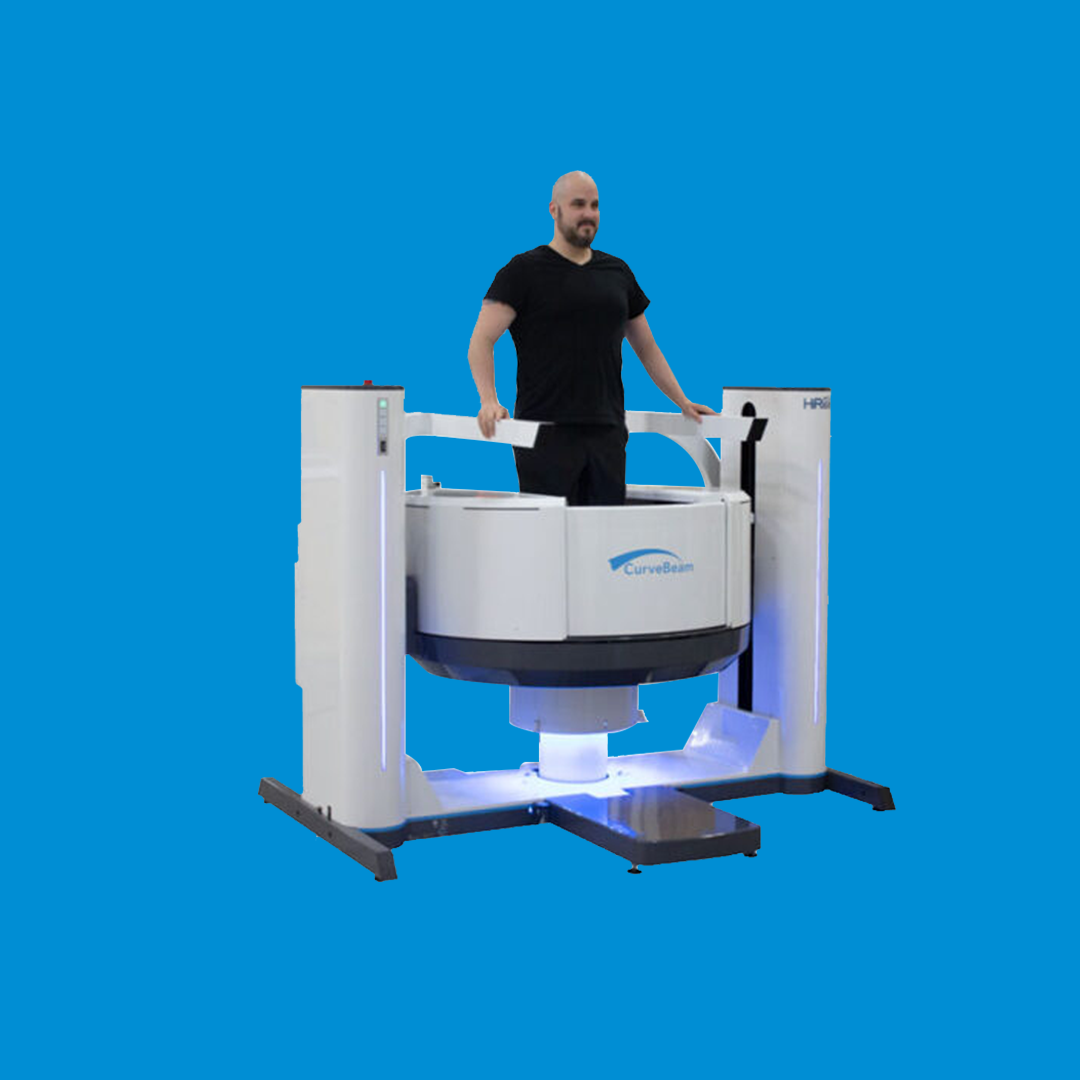
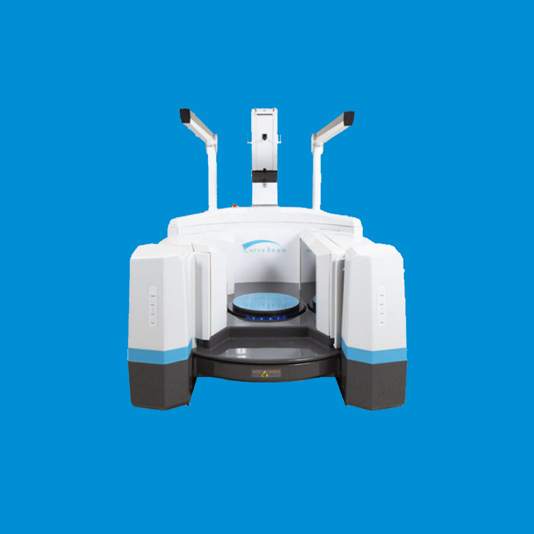
InReach is an advanced CT imaging product from CurveBeam AI. It offers point-of-care extremity CT imaging for the upper extremities and non-weight bearing lower extremities. Specialists can use the data from InReach to assess osseous structures and plan procedures accordingly. The level of precision and clarity that comes from InReach helps improve operational efficiency within hospitals and clinics. It also helps the relationship between orthopaedic distributors and hospitals, as the latter have a clearer idea of the implants they need.
Aid Pre-Op Planning
Medical professionals mainly use the InReach system to aid pre-op planning. This can include complications associated with the hand, wrist forearm, elbow, and lower extremities like the ankle. Experts have reported InReach has been consistently useful for distal radius fractures, scaphoid fractures, midcarpal fusions and more.
A further benefit of InReach is the low dose of radiation that comes with its scans. One InReach scan imparts an effective radiation dose of around 1 micro sievert, where the average person in the US receives roughly 3000 micro sieverts per year from background sources. This low dosage is thanks to the use of Cone Beam CT scanning, which is employed across the CurveBeam AI range. For InReach, the default voxel size is 0.2mm. CBCT is therefore a viable alternative for paediatric patients that would otherwise receive a conventional medical CT (MDCT).
The InReach system can be installed easily due to its small dimensions:
Interested in InReach
If you would like more information regarding InReach

The Standing CT Company is a healthcare supplier committed to improving patient experiences through the provision of industry-leading technology. Their primary focus in on accurate 3D imaging of the lower and upper extremities for use by orthopaedic clinicians, as well as the core values of ethics and sustainability. The Standing CT Company partners with other experts to deliver these quality services, namely CurveBeam AI.
CurveBeam AI offers proprietary tools for hospitals to improve patient outcomes. The company combines market leading point-of-care diagnostic cone beam CT imaging solutions, with artificial intelligence (AI) and deep learning AI (DLAI) expertise. This allows CurveBeam AI to deliver fragility fracture prevention solutions for bone health across many areas of orthopaedics. Weight bearing CT imaging is a core focus for the business, alongside building complementary AI tools that allow solutions to bridge disease care.
It’s only when individual doses exceed 100 micro sieverts that radiation poses a risk to public health. Consistent exposure to low levels of radiation is natural for members of the public to experience. An InReach, scan doesn’t represent a drastic increase from these levels with just a 1 micro sieverts dose. For context, a traditional CT scan of the unilateral foot and ankle will cause an exposure of 25-70 micro sieverts.
A CT scanner collects image data by rotating 360 degrees. Multiple rotations are needed for an MDCT to capture an area due to the narrow fan-shaped beam it uses. A CBCT, on the other hand, will instead rotate once to acquire image projections. These are then reconstructed to into a series of axial slices. The CBCT plat panel detector is positioned closer to patient anatomy than in a MDCT.