4th July 2025
Mersey 58 Society Registrar’s Day
Isla Gladstone Conservatory, L4 0TD
Features
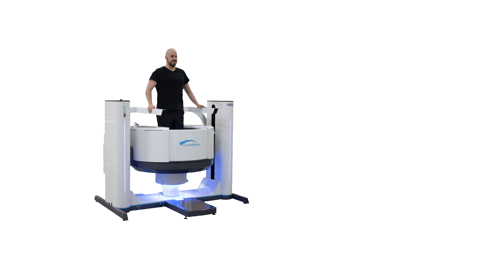
HiRise is a bilateral weight bearing CT imaging device. It offers multi-extremity solutions, able to produce detailed computer tomography scan images of the hips, knees, foot, ankles, and upper extremities. The system features a CT image detector and a patient chair.
HiRise follows a protocol based on the part of the body it is being used to image. All of these bar the MDCT hip protocol, deliver a dose below the average background level of radiation. The system also features a flexible gantry that allows for easy positioning to scan upper extremities. This is particularly useful for patients who are wheelchair bound or unable to stand.
Manoeuvrable and Compact Design
The HiRise chair can accommodate up to 204kg. Its design is manoeuvrable and compact, thus allowing for simple storage when the system is not in use.
CT scan technology creates detailed 3D images of internal body structures like bones, organs and blood vessels. Orthopaedic Conebeam CT is specialised to scan bones and weight-bearing allows surgeons to see joint spacing and bone positioning while the patient is standing.
Cone Beam CT Imaging vs Regular CT Imaging
Cone beam CT scanners use a medical fluoroscopy tube to generate radiographs. Traditional CT scanners, on the other hand, use anode x-ray tubes which produce far higher amounts of radiation. This is why HiRise can capture high resolution images while generating low levels of radiation. As you might guess, CBCT scans send x-rays out in a cone shaped beam. For medical imaging, conventional CT scans will use either a fan or spiral shaped beam.
HiRise Cone Beam CT images are initially acquired as two-dimensional projections, using a rotating gantry with a fixed-anode X-Ray tube ring, a pulsed X-Ray beam, and a flat panel detector. The gantry rotates 360° to acquire image projections which are then reconstructed using a series of axial slices.
Interested in HiRise
If you would like more information regarding HiRise
Specifications


The Standing CT Company is a healthcare supplier committed to improving patient experiences through the provision of industry-leading technology. Their primary focus in on accurate 3D imaging of the lower and upper extremities for use by orthopaedic clinicians, as well as the core values of ethics and sustainability. The Standing CT Company partners with other experts to deliver these quality services, namely CurveBeam AI.
CurveBeam AI offers proprietary tools for hospitals to improve patient outcomes. The company combines market leading point-of-care diagnostic cone beam CT imaging solutions, with artificial intelligence (AI) and deep learning AI (DLAI) expertise. This allows CurveBeam AI to deliver fragility fracture prevention solutions for bone health across many areas of orthopaedics. Weight bearing CT imaging is a core focus for the business, alongside building complementary AI tools that allow solutions to bridge disease care.
For lower limb surgeons, being able to see the joint spacing and bone positioning in a load-bearing, functional position is very important. Through the use of Conebeam CT, 3D weight-bearing scans are available at a radiation dosage that is roughly equivalent to the corresponding X-ray set. Traditional weight-bearing X-rays can be problematic as they can take time to position the patient correctly and multiple X-rays are required which takes time and is not consistently reproducible. With HiRise standing CT scans, the contralateral limb is also always imaged at the same time, as a control.
A standing CT is useful in almost all clinical cases where a weight-bearing X-ray or CT is traditionally indicated.
Cone Beam CT (CBCT) uses a concentrated, low-power X-ray beam instead of a fan-shaped, high-output X-ray beam. This results in a lower radiation dosage and means a CBCT is much quicker than traditional “slice” CT. In addition both legs are scanned at the same time, allowing the opposite side to act as a comparison without you having to have another set of scans taken. This also removes the need for additional radiation exposure.
Yes. The HiRise produces 3D alignment scans in a weight-bearing position that allows lower limb surgeons to see the full alignment from hips to toes.
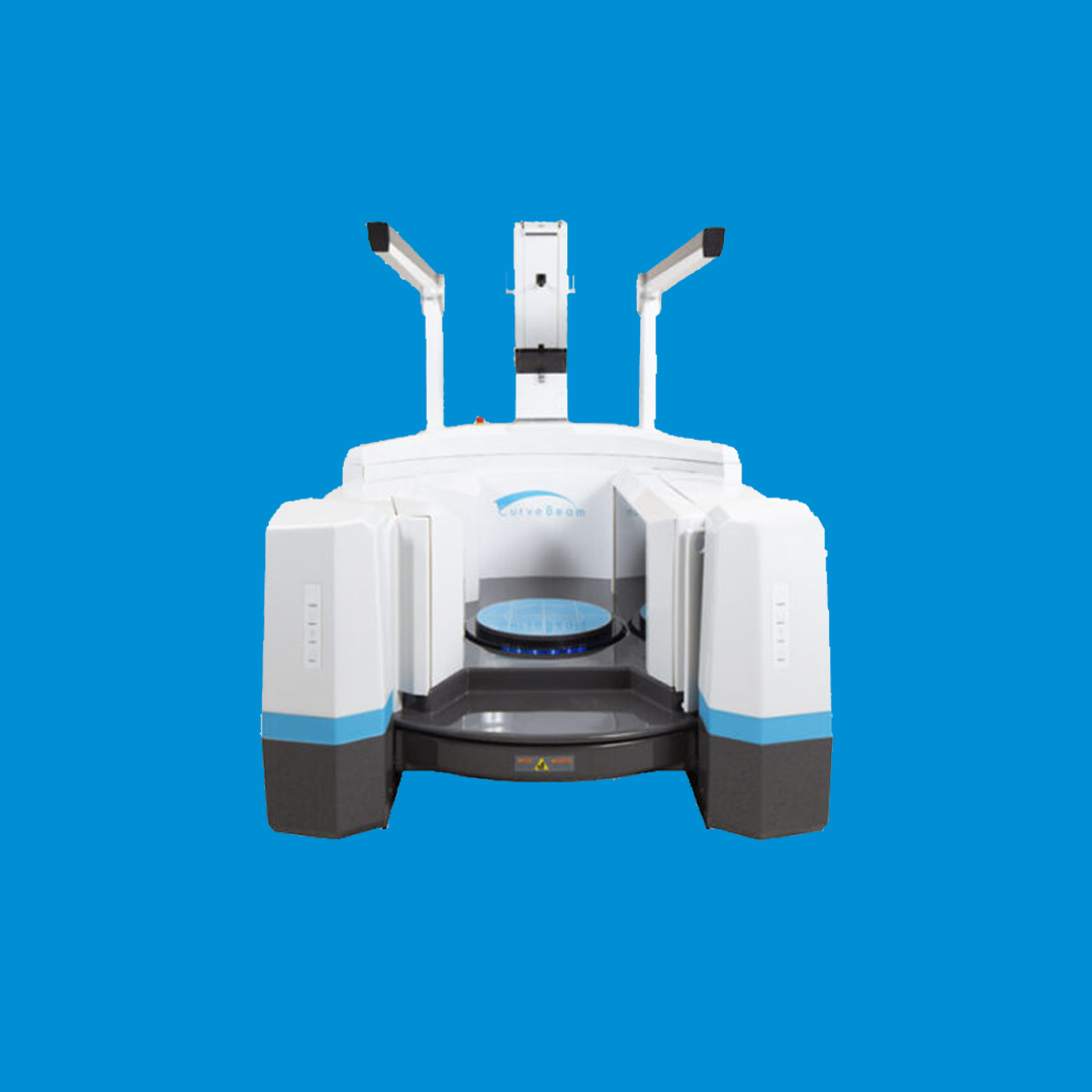
The HiRise scanner reliably images both feet and ankles, knees or hips in a single pass without the patient having to change their simple normal standing position. This gives referrers a true bilateral comparison. Other scanners scan the forefoot or hindfoot separately and then use a software algorithm to stitch the images together.
Scans are also quick (30-60 seconds) and deliver a low radiation dosage. Our scanners have a walk-in access port and, where required, a seat and handrail. This means that elderly patients or patients in a plaster can be scanned more easily than with equipment which require the patient to climb up or into the scanner.
The Conebeam CT scanners from the Standing CT Company allow rapid 3D images at very low radiation dosage compared to conventional CT. For a scan of the upper limb, the patient simply puts their hand/wrist or elbow into the scanner (rather than having to be scanned in a superman position, as they do with conventional CT)